
Ultrasound imaging, also known as sonography, is a non-invasive diagnostic technique that uses high-frequency sound waves to create images of structures inside the body. During pregnancy, ultrasound scans are commonly used to monitor the development and well-being of the fetus.
Here are key points to understand about ultrasounds during pregnancy
1. Purpose: Ultrasound scans are performed at various stages of pregnancy to assess fetal growth, anatomy, and position, as well as to monitor the placenta and amniotic fluid levels. They help healthcare providers evaluate the health of the fetus and detect any potential abnormalities.
2. Timing: Ultrasound scans are typically performed at specific timepoints during pregnancy:
Dating Scan (First Trimester): This ultrasound is performed between 8 to 14 weeks of pregnancy to confirm the due date, assess the number of fetuses, and check for the presence of a heartbeat.
Nuchal Translucency (NT) Scan (First Trimester): Conducted between 11 to 14 weeks, this scan measures the thickness of fluid at the back of the baby’s neck to assess the risk of chromosomal abnormalities.
Anomaly Scan (Second Trimester): This detailed ultrasound is performed around 18 to 22 weeks to evaluate the baby’s anatomy and detect any structural abnormalities.
Growth Scan (Third Trimester): If necessary, additional ultrasound scans may be performed in the third trimester to monitor fetal growth and well-being, especially in cases of high-risk pregnancies.
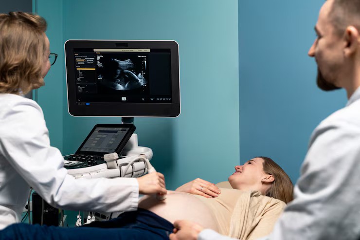
3. Procedure: During an ultrasound scan, a gel is applied to the mother’s abdomen, and a handheld device called a transducer is moved over the gel-covered area. The transducer emits sound waves that bounce off internal structures, creating images that are displayed on a monitor. In some cases, transvaginal ultrasounds may be performed in early pregnancy for better visualization.
4. Safety: Ultrasound imaging is considered safe and does not use ionizing radiation, making it suitable for routine prenatal screening. However, it’s essential to limit unnecessary exposure to ultrasound scans and only undergo them when recommended by a healthcare provider.
5. Interpretation: The images generated by ultrasound scans are interpreted by trained professionals, such as radiologists or obstetricians. They assess various aspects of fetal development, including the size and anatomy of organs, the position of the placenta, and the volume of amniotic fluid.
Ultrasound scans play a crucial role in prenatal care, providing valuable information about the health and development of the fetus. It’s essential to discuss the purpose and findings of ultrasound scans with your healthcare provider to ensure the best possible pregnancy outcomes. If you have any questions or concerns about ultrasound imaging during pregnancy, don’t hesitate to reach out to your healthcare provider for guidance and support.